What a bone stress injury actually is
Bone is living tissue. Every run bends it a tiny amount, and those loading cycles leave behind microscopic damage. Normally the body removes that damage and lays down new bone, and over months the skeleton gets stronger for the running you do. A bone stress injury happens when damage builds faster than repair can keep up (Hoenig et al., 2022a).
It sits on a spectrum. Early on, MRI shows swelling in the bone or its lining with no visible break; that is a stress reaction. Further along, a fracture line appears. Clinicians now prefer the umbrella term "bone stress injury" because most runners I see are somewhere in the middle, not at either end.
Risk factors, symptoms, and signs
The most common story I hear is a jump in training. A new coach, a return after time off, a marathon block that added mileage and speed work at the same time. When the number and size of loading cycles outpace what the bone can tolerate, injury follows (Warden et al., 2021). Training load is rarely the only factor, though. A prior bone stress injury is one of the strongest red flags: in one college cohort, 50 to 63% of athletes with a new injury had a previous one, compared with 11% of uninjured athletes (Tenforde et al., 2022). Female runners are affected more often than males, and low energy availability, menstrual disruption, and low bone density raise risk in runners of any sex (Hoenig et al., 2022a).
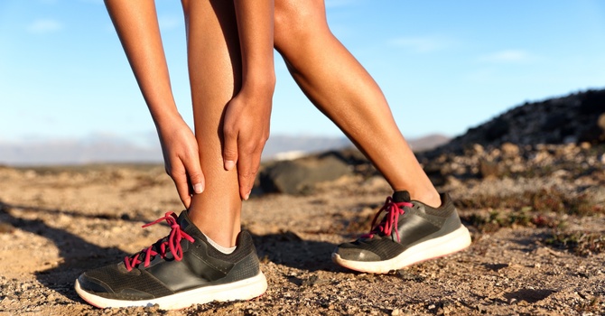
Symptoms usually build gradually. Early on, pain shows up late in a run and settles with rest. Over weeks it arrives sooner, lingers after the run, and eventually hurts with walking or at night. On exam, the classic sign is tenderness you can point to with one finger over the bone itself. Pain with single-leg hopping supports the diagnosis (Hoenig et al., 2022a). A diffuse ache spread along several inches of the shin is more typical of medial tibial stress syndrome, but the two overlap, and I treat that line as blurry rather than fixed.
How bone stress injuries are tested
The clinical exam gets us most of the way to a working diagnosis: the training history, focal bony tenderness, and pain with hopping. Bedside tools have been tried as shortcuts. A meta-analysis of tuning fork and therapeutic ultrasound tests found ultrasound had a pooled sensitivity and specificity of only about 64% and 63%, and the authors concluded neither should be used on its own to diagnose a stress fracture (Schneiders et al., 2012).
Imaging confirms the diagnosis. Plain X-rays are often normal in the first weeks, so a clean X-ray does not rule anything out. MRI is far more sensitive for early injury (Hoenig et al., 2022a), and it grades severity, from swelling in the bone lining (grade 1) up to a visible fracture line (grade 4). That grade matters because it predicts how long recovery will take, which I cover below. When we suspect a high-risk site, we push for imaging early rather than waiting to see how it feels.
Management: nutrition, rest, strength, and time
Where the injury is changes everything. Most runners have a low-risk injury in bone that is loaded mainly in compression, like the back of the shin or the middle metatarsals. These heal reliably with modified activity and a graded return to loading. High-risk sites, such as the tension side of the femoral neck, the navicular, the front of the tibia, and the base of the fifth metatarsal, carry a real chance of delayed healing or a complete fracture. They may need a boot, crutches, or surgical consultation (Hoenig et al., 2022a).
Rest means relative rest, not bed rest. For low-risk injuries, we stop running and keep everything that is pain-free: walking, cycling, pool running, and gym work. Pain is the guide. Daily activity should be pain-free before any running starts, and running comes back as walk-run intervals on alternate days so the bone recovers between sessions.
Fuel is part of the treatment. Bone cannot rebuild without enough energy. The International Olympic Committee now uses the term Relative Energy Deficiency in Sport (REDs) for the health effects of eating too little for the training you do, and it applies to men as well as women (Mountjoy et al., 2023). Athletes with menstrual disruption tend to have higher-grade injuries, and lower bone density predicts a slower return (Nattiv et al., 2013). If a runner has had more than one bone stress injury, a sports dietitian and a physician belong on the team.
Strength training does more than keep you fit. Stronger calf, quad, and hip muscles help absorb the forces of running, and loaded, varied movement is how bone gets stronger in the first place (Warden et al., 2021). We start early with exercises that don't hurt, then progress to single-leg strength and later to hopping and landing before a full return. Small running changes also help, such as a slightly higher cadence, which reduces the load on bone with each step (Warden et al., 2021).
How long it takes depends on the MRI grade. A meta-analysis pooled the average time to return to sport by grade (Hoenig et al., 2022b):
|
MRI grade |
Average return to sport |
|---|---|
|
Grade 1 |
42 days (about 6 weeks) |
|
Grade 2 |
70 days (about 10 weeks) |
|
Grade 3 |
84 days (about 12 weeks) |
|
Grade 4 |
99 days (about 14 weeks) |
High-grade injuries in trabecular-rich bone, such as the femoral neck, pelvis, and sacrum, added nearly twice as much time as those in cortical bone like the tibia. The good news is that more than 90% of athletes returned to sport (Hoenig et al., 2022b).
The limits of screening
Runners often ask whether a test can tell them they are at risk before anything hurts. The honest answer is not reliably. A risk factor that is more common in injured groups does not become a useful test for an individual, because the ranges for injured and uninjured athletes overlap heavily. As one widely cited review put it, there is no screening test for sports injury with adequate predictive properties (Bahr, 2016).
Imaging healthy runners doesn't solve this either. In one study, 43% of asymptomatic college distance runners had signs of a tibial stress reaction on MRI, and those findings did not predict who later developed a symptomatic injury (Bergman et al., 2004). Questionnaires that add up energy-availability and menstrual risk factors do track with injury rates across groups (Tenforde et al., 2022), and they are worth using to start a conversation. They still can't tell any one runner whether they will get hurt. So we use screening to find modifiable problems like underfueling, missing periods, or a sudden spike in mileage, and fix them. We don't use it to promise anyone they are safe.
When to get it checked
If you have bone pain you can point to with one finger, pain that has started to show up with walking, or pain that hurts when you hop or jump, stop running and get evaluated. Catching a stress reaction at grade 1 usually means about six weeks away from running. Pushing through it can turn it into a fracture and a season.
At OSO, every evaluation is one-on-one with a board-certified orthopedic specialist. We look at your training history, your strength, and how you run, and we coordinate with your physician when imaging is needed. Then we build a return-to-run plan. If you're a runner in Alameda or the East Bay dealing with shin, foot, or hip pain that isn't going away, contact us to schedule an evaluation.
References
- Bahr, R. (2016). Why screening tests to predict injury do not work—and probably never will…: A critical review. British Journal of Sports Medicine, 50(13), 776–780. https://doi.org/10.1136/bjsports-2016-096256
- Bergman, A. G., Fredericson, M., Ho, C., & Matheson, G. O. (2004). Asymptomatic tibial stress reactions: MRI detection and clinical follow-up in distance runners. American Journal of Roentgenology, 183(3), 635–638. https://doi.org/10.2214/ajr.183.3.1830635
- Hoenig, T., Ackerman, K. E., Beck, B. R., et al. (2022a). Bone stress injuries. Nature Reviews Disease Primers, 8, 26. https://doi.org/10.1038/s41572-022-00352-y
- Hoenig, T., Tenforde, A. S., Strahl, A., Rolvien, T., & Hollander, K. (2022b). Does magnetic resonance imaging grading correlate with return to sports after bone stress injuries? A systematic review and meta-analysis. The American Journal of Sports Medicine, 50(3), 834–844. https://doi.org/10.1177/0363546521993807
- Mountjoy, M., Ackerman, K. E., Bailey, D. M., et al. (2023). 2023 International Olympic Committee's (IOC) consensus statement on Relative Energy Deficiency in Sport (REDs). British Journal of Sports Medicine, 57(17), 1073–1097. https://doi.org/10.1136/bjsports-2023-106994
- Nattiv, A., Kennedy, G., Barrack, M. T., Abdelkerim, A., Goolsby, M. A., Arends, J. C., & Seeger, L. L. (2013). Correlation of MRI grading of bone stress injuries with clinical risk factors and return to play: A 5-year prospective study in collegiate track and field athletes. The American Journal of Sports Medicine, 41(8), 1930–1941. https://doi.org/10.1177/0363546513490645
- Schneiders, A. G., Sullivan, S. J., Hendrick, P. A., et al. (2012). The ability of clinical tests to diagnose stress fractures: A systematic review and meta-analysis. Journal of Orthopaedic & Sports Physical Therapy, 42(9), 760–771. https://doi.org/10.2519/jospt.2012.4000
- Tenforde, A. S., Katz, N. B., Sainani, K. L., Carlson, J. L., Golden, N. H., & Fredericson, M. (2022). Female athlete triad risk factors are more strongly associated with trabecular-rich versus cortical-rich bone stress injuries in collegiate athletes. Orthopaedic Journal of Sports Medicine, 10(9), 23259671221123588. https://doi.org/10.1177/23259671221123588
- Warden, S. J., Edwards, W. B., & Willy, R. W. (2021). Preventing bone stress injuries in runners with optimal workload. Current Osteoporosis Reports, 19(3), 298–307. https://doi.org/10.1007/s11914-021-00666-y

Ben Fedewa
Contact Me