Knee pain after activity is one of the most common things I see in my practice. Someone goes for a run, comes home and feels fine, and wakes up the next morning with an ache that was not there the day before. Or they start a new training program, feel great for a few weeks, and then the knee starts complaining during squats or on stairs. Sometimes it comes on during the activity itself, a dull burn around the kneecap that builds with mileage or load. Most people assume the knee is broken in some way. They want an MRI, or they want to know if their cartilage is worn down, or they are convinced they have done something structurally significant. And sometimes that is true. But far more often, what I find when I evaluate someone with activity related knee pain is that the knee is actually the most innocent joint in the room. It is absorbing forces it was never supposed to absorb because something above it or below it is not doing its job. Knee pain is genuinely common. The annual prevalence of patellofemoral pain alone, which is one of the most frequent presentations of activity related anterior knee pain, has been reported at 22.7 percent in the general population and as high as 28.9 percent among adolescents (Smith et al., 2018). A separate survey of adults aged 18 to 39 found that nearly one in three reported knee problems in the past year, with pain being the dominant complaint in 65 percent of cases (Smith et al., 2019). These are not rare or unusual numbers. Knee pain affects a huge portion of the physically active population. What I want to do in this post is explain how I think about it, because the way most people understand knee pain and the way it actually works are often very different. The knee sits in the middle of a chain. What happens at the foot and ankle and what happens at the hip and pelvis have direct, measurable consequences for how much stress the knee is under during every single step you take. When we fix the knee without addressing those factors, we get short term relief at best. When we address the whole chain, people get better and stay better. The knee is almost never the only problem. It is usually where the chain breaks down, but the chain starts at the foot and runs all the way to the hip. That is where the evaluation has to begin.
The Phases of Knee Pain
Just as with back pain, where someone is in the timeline of their knee problem matters a great deal for how I approach treatment. Trying to load a knee that is in the middle of an acute inflammatory episode is counterproductive. Treating a chronic knee problem with only pain relief strategies without building real capacity is how people end up back in the clinic six months later. The phases are distinct, and the treatment in each one looks different. Phase 1: Calming Things Down When knee pain flares, whether from a spike in training volume, a long hike, or just a bad day at the gym, the first priority is getting the irritability under control. The joint is inflamed, and in that state it is not going to respond well to loading. Pushing through significant pain to train is not toughness; it is tissue damage accumulation, and it sets the rehab timeline back.
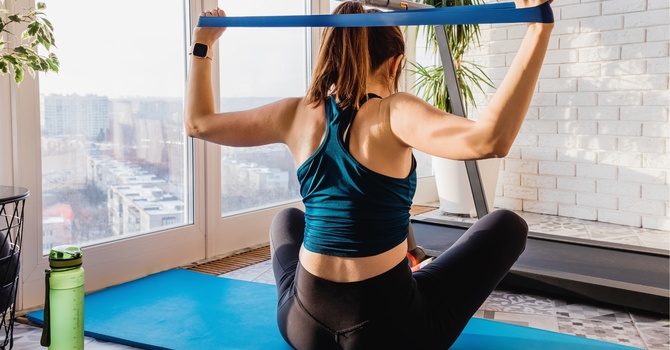
Phase 1 : I am focused on a few things. First, load management. We identify what activities are driving symptoms and temporarily modify them. This does not mean stopping everything. It usually means pulling back on the specific demands that are stressing the joint, whether that is running volume, squatting depth, or stair use, and replacing them with lower load alternatives that keep the body moving without irritating the tissue. Second, manual therapy. Targeted joint mobilization, soft tissue work to the quadriceps, IT band complex, and calf, and sometimes patellar mobilization can meaningfully reduce pain and restore movement in a knee that has locked up due to guarding and swelling. This hands on work is not a cure, but it lowers the pain level enough that the patient can start moving well again, which is the foundation for everything that comes next. Third, neuromuscular activation. Even in the pain control phase I want to start waking up the muscles that have gone quiet around the joint, specifically the Quads, the gluteus medius, and the deep hip external rotators. These muscles shut down quickly in the presence of pain and swelling (arthrogenic and neurogenic inhibition), and the longer they are irritated the harder they are to recruit later. Gentle isometric and low load work begins here, possibly with BFR or NMES.
Phase 2: Once the acute flare has settled and the patient can move without significant pain, we get into the real work. This is the phase where we figure out why the knee got irritated in the first place, because the answer is almost never found at the knee itself. In my evaluation I am looking at three areas: the hip, the knee locally, and the foot and ankle. All three have to be examined systematically, because the sources of dysfunction can be anywhere in that chain and they often interact with each other. What follows is how I think about each one.
What the Hip Has to Do With It
The hip is the most important joint to examine in any patient with activity related knee pain, and it is also the most commonly overlooked. The reason is biomechanical and pretty straightforward once you understand it. During every weight bearing activity, from walking to running to squatting to landing from a jump, the femur rotates under the patella. If the hip abductors and external rotators are strong and firing properly, they control that rotation. The femur stays in a relatively neutral position, the patella tracks cleanly through the trochlear groove, and the joint loads are distributed evenly. When those muscles are weak or faulty in their timing, the femur rotates inward and adducts, which drives the knee into a valgus position and lateralizes the patellar contact point, concentrating stress on tissues that are not designed to handle it. The research on this is very consistent. A systematic review with meta-analysis published in Healthcare (Shu et al., 2022) found strong evidence that individuals with patellofemoral pain have significantly weaker hip external rotators and hip abductors compared to pain free controls. A prior systematic review in the Journal of Orthopaedic and Sports Physical Therapy (Powers, 2010) established the biomechanical mechanism clearly: impaired muscular control of the hip, pelvis, and trunk alters both tibiofemoral and patellofemoral kinematics in multiple planes, contributing to conditions ranging from patellofemoral pain to IT band syndrome. Prospective data makes it even more compelling. Research by Noehren and colleagues found that runners who went on to develop patellofemoral pain demonstrated greater peak hip internal rotation during running than those who remained pain free, suggesting that the faulty hip mechanics preceded the knee pain rather than being a consequence of it. Just 10 degrees of femoral internal rotation has been shown to substantially decrease patellofemoral joint contact area and increase joint stress by as much as 50 percent (Powers, 2003). A systematic review and meta-analysis in the Journal of Orthopaedic and Sports Physical Therapy by Nascimento and colleagues (2018) put the clinical question to rest as well as anything in the literature: combining hip strengthening with knee strengthening is more effective than knee strengthening alone for reducing pain and improving activity in people with patellofemoral pain. Of 14 randomized controlled trials involving 673 participants, the combined approach consistently outperformed isolated knee treatment. Hip and knee work together. Treating only one of them is leaving results on the table. In clinical practice this means that whenever I see a patient with activity related knee pain, I am testing hip abductor and external rotator strength, watching their single leg squat mechanics, checking for a Trendelenburg pattern during gait, and assessing their ability to control femoral rotation under load. Those findings drive the exercise prescription, not just where it hurts. Strengthening the hip in combination with the knee is more effective than knee strengthening alone for reducing pain in patellofemoral pain patients. Treating only the knee is treating only part of the problem.
What the Foot and Ankle Have to Do With It
If the hip is the most overlooked joint in knee pain evaluation, the foot and ankle are a close second. This one surprises people more, because it feels like a longer leap from the foot to the kneecap. But the chain is continuous, and what happens at ground contact ripples up through every joint above it. There are two main things I look at in the foot and ankle: ankle dorsiflexion range of motion and foot pronation patterns. Both of them can independently drive knee pain, and in many patients they interact with each other. Ankle Dorsiflexion Ankle dorsiflexion is the ability of the ankle to bend as the shin moves forward over the foot. You need it for squatting, for stair descent, for running, and for essentially any athletic movement that requires the knee to flex under load. When dorsiflexion is restricted, whether because of a tight gastrocnemius and soleus complex, joint capsule stiffness from an old ankle sprain, or bone morphology, something upstream has to compensate. The research on this is clear and clinically significant. A study by Dill and colleagues (2014), published in the Journal of Athletic Training, found that individuals with limited weight bearing lunge ankle dorsiflexion range of motion demonstrated significantly altered knee and ankle kinematics during squatting, with reduced knee flexion angles and increased knee valgus. When the ankle cannot dorsiflex adequately to allow the shin to travel forward, the knee compensates by collapsing inward, and the foot compensates by pronating excessively at the subtalar joint. Both of those compensation patterns increase patellofemoral joint stress. A 2022 study published in Frontiers in Physiology confirmed that restricted ankle dorsiflexion is associated with increased frontal plane knee motion during landing and squatting tasks, and that athletes with restricted dorsiflexion tend to display a stiff legged landing pattern rather than properly absorbing load through knee and hip flexion (Kerin and colleagues, 2022). Stiff legged landings are associated with greater vertical ground reaction forces and tibiofemoral compression, which translate directly into more stress on the knee. In practice this means that when I evaluate a patient with anterior or peripatellar knee pain, I always assess dorsiflexion with a weight bearing lunge test. A restriction of less than roughly 10 centimeters of forward knee travel past the toes with the heel flat on the floor tells me there is likely a mobility deficit contributing to the problem. Addressing it, whether through manual mobilization of the talocrural joint, soft tissue work to the posterior chain, or progressive calf stretching with load, often produces an immediate reduction in symptoms during functional movements. Foot Pronation and the Kinetic Chain Foot pronation is a normal and necessary part of gait. The subtalar joint pronates at initial contact to absorb shock, and then supinates through midstance and pushoff to create a rigid lever for propulsion. The problem arises when the foot stays pronated too long or excessively, because prolonged subtalar pronation prevents the tibia from externally rotating as the limb extends. When that happens, the femur compensates with internal rotation, which drives the familiar pattern of knee valgus and lateral patellar tracking stress. A systematic review published in the Journal of Clinical Medicine (Rabelo and colleagues, 2022) examined the relationship between foot and ankle alignment and patellofemoral pain syndrome, finding that a more pronated foot posture was associated with PFPS in several studies, with one reporting significantly greater foot mobility and pronation in the PFPS group compared to controls. A narrative review in Frontiers in Physiology (Mei and colleagues, 2022) confirmed that overpronated foot posture is associated with internally rotated tibia, increased patellar mobility, and anterior knee pain during running. What this tells me clinically is that in a patient whose knee pain is provoked by running or repetitive lower extremity loading, I need to watch how their foot contacts the ground and whether they have the intrinsic foot strength and ankle mobility to control pronation through the loading phase. Flat feet in isolation are not a problem. Flat feet combined with weak hip external rotators, poor ankle mobility, and inadequate intrinsic foot strength create a situation where the knee has no protection from the forces being transmitted up from the ground. Treatment in this area often includes intrinsic foot strengthening, calf and posterior chain mobility work, gait or movement pattern retraining, and sometimes a discussion about footwear or temporary orthotics if the structural demands are beyond what strengthening can address in a reasonable time frame.
Phase 3: Building Capacity and Staying Out of the Clinic
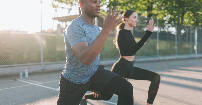
Phase 3 for the knee is the same concept as it is for the back: systematically reloading the joint in a way that builds real tolerance to the demands the patient is asking of it. The difference between someone who fully recovers from knee pain and someone who manages it chronically is almost always whether they completed this phase. Completing Phase 2 gets the pain down and addresses the biomechanical contributors. But that does not mean the knee is ready to handle full training volumes, maximum loading, or sport specific demands. The tissues need progressive loading to adapt, and the neuromuscular system needs to practice the movement patterns it learned in controlled conditions under the kind of speed, fatigue, and unpredictability that actually matches real life.
Phase 3 at OSO PT typically includes:
• Progressive lower extremity strength work: squatting, lunging, deadlifting, and step down progressions loaded to a level that challenges the tissue without irritating it
• Single leg loading: a significant focus because the demands of running and most sports are almost entirely single limb • Plyometric and reactive training: for patients returning to sport, the ability to absorb and redirect force through the knee quickly and under fatigue is a specific skill that has to be trained, not assumed
• Running gait or sport specific mechanics: if someone is a runner, I want to see them run. Addressing form faults in step rate, trunk lean, foot strike pattern, or arm carry can meaningfully reduce knee load without requiring any strength change at all • Load management planning: building a sustainable return to full training with enough structure to prevent the next spike in symptoms The goal is not just a pain free knee. It is a knee that can handle everything the patient wants to do, consistently, without needing to come back in every few months for a tune up.
Why This Keeps Coming Back
The number one reason knee pain is recurrent is that the root cause was never adequately addressed. The treatment stayed local to the knee: ice, quad sets, some leg extensions, maybe an IT band stretch, and the patient felt better enough to go back to training. Then a few months later the same thing happens again. What was missed is almost always the same list: hip abductor and external rotator endurance under fatigue, dorsiflexion mobility that never got restored after an old ankle sprain, foot mechanics that have been compensating for years. These are not things that resolve on their own. They require specific, targeted work, and they require enough progressive loading that the improvements hold up under real world demands. When I evaluate someone who has had knee pain on and off for years, the evaluation findings are usually not subtle. The hip weakness is significant. The dorsiflexion restriction is measurable. The single leg squat mechanics are poor. These things did not develop because of bad luck. They developed over time, and they will return if the treatment does not actually fix them.
When to Come In
You should consider seeing a Board Certified Orthopaedic Physical Therapist about your knee pain if any of the following apply: • Pain that lingers after activity for more than 24 to 48 hours
• Pain that is getting progressively worse with training rather than adapting
• Swelling or a feeling of instability in the joint
• Pain that is limiting your ability to run, squat, use stairs, or do the activity you care about
• Recurring episodes that seem to resolve and then come back with the next training cycle
• You have been told to rest and the rest is not fixing anything
In California you can see a physical therapist directly without a referral. You do not need to wait, and you do not need a diagnosis in hand to start the evaluation. Getting an accurate picture of what is actually driving your pain is the most important first step, and it often produces results faster than people expect.
From the Clinic
Knee pain is extremely common, but it does not have to be permanent, and it definitely does not have to keep coming back. In my experience, the vast majority of patients with activity related knee pain can get back to full function when the evaluation is thorough enough to find the actual problem and the treatment is progressive enough to build real capacity. If you are running through knee pain, modifying your training to work around it, or dealing with the same problem for the third or fourth time, come in. We will do a full evaluation, look at the whole chain, find what is actually driving it, and build a plan that addresses it at the source. Call or text at 510-915-1448, or email dan.hirai@osophysicaltherapy.com. Free phone screens are always available if you want to talk through what you are dealing with before scheduling.
References
Dill, K. E., Begalle, R. L., Frank, B. S., Zinder, S. M., and Padua, D. A. (2014). Altered knee and ankle kinematics during squatting in those with limited weight bearing lunge ankle dorsiflexion range of motion. Journal of Athletic Training, 49(6), 723 732. Kerin, F., Farrell, G., Tierney, A., McCarthy Persson, U., De Vito, G., and Delahunt, E. (2022). Ankle dorsiflexion affects hip and knee biomechanics during landing. International Journal of Sports Physical Therapy, 17(3), 366 376. Macrum, E., Bell, D. R., Boling, M., Lewek, M., and Padua, D. (2012). Effect of limiting ankle dorsiflexion range of motion on lower extremity kinematics and muscle activation patterns during a squat. Journal of Sport Rehabilitation, 21(2), 144 150. Mei, Q., Gu, Y., Xiang, L., Baker, J. S., Fernandez, J., and Ugbolue, U. C. (2022). Toward improved understanding of foot shape, foot posture, and foot biomechanics during running: A narrative review. Frontiers in Physiology, 13, 1062598. Nascimento, L. R., Teixeira-Salmela, L. F., Souza, R. B., and Resende, R. A. (2018). Hip and knee strengthening is more effective than knee strengthening alone for reducing pain and improving activity in individuals with patellofemoral pain: A systematic review with meta-analysis. Journal of Orthopaedic and Sports Physical Therapy, 48(1), 19 31. Powers, C. M. (2010). The influence of abnormal hip mechanics on knee injury: A biomechanical perspective. Journal of Orthopaedic and Sports Physical Therapy, 40(2), 42 51. Powers, C. M. (2003). Patellar kinematics, part I: The influence of vastus muscle activity in subjects with and without patellofemoral pain. Physical Therapy, 83(12), 1071 1082. Rabelo, N. D. de A., Dos Santos Araujo, T., Oliveira, A. S., Lucareli, P. R. G., and Lahoz, G. L. (2022). Does the foot and ankle alignment impact the patellofemoral pain syndrome? A systematic review and meta-analysis. Journal of Clinical Medicine, 11(8), 2245. Shu, Y., and colleagues. (2022). The relationship between patellofemoral pain syndrome and hip biomechanics: A systematic review with meta-analysis. Healthcare, 11(1), 99. Smith, B. E., Selfe, J., Thacker, D., Hendrick, P., Bateman, M., Moffatt, F., Corbett, M., Logan, P., and Doyle, C. (2018). Incidence and prevalence of patellofemoral pain: A systematic review and meta-analysis. PLOS ONE, 13(1), e0190892. Smith, T. O., Ooms, A., Smith, B., and Hing, C. B. (2019). Knee problems are common in young adults and associated with physical activity and not obesity: The findings of a cross-sectional survey in a university cohort. BMC Musculoskeletal Disorders, 20(1), 112.

Ben Fedewa
Contact Me